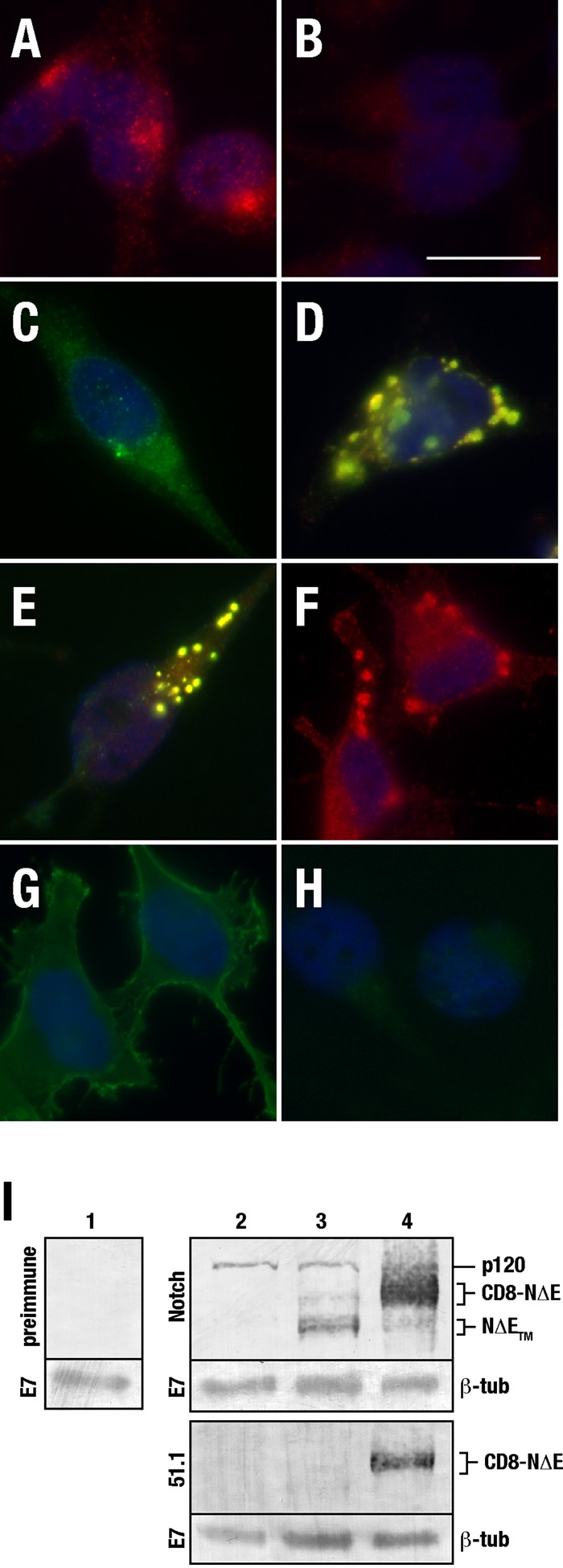
FIGURE 1:

Notch immunolocalization analysis. tTA HeLa cells were fixed and probed with antisera against NICD (A) or preimmune sera (B). tTA HeLa cells were transfected with plasmid encoding CD8-mNotch under control of the tetracycline-regulatable (C, pTREL , low expression levels) or CMV promotor (D, pCMV, high expression levels), fixed, and probed for the CD8 epitope using mAb 51.1 (green) or NICD (red). tTA HeLa cells were infected with adenovirus encoding CD8-NΔE (E), NΔE-6myc (F), or CD8-LDLR (G), and probed with 51.1 (green) and/or NICD (red). (H) 51.1 antibody background. Yellow indicates overlap of the red and green channels. (I) Immunoblot analysis of control cells (lanes 1 and 2) or those expressing pTREL-CD8-NΔE (lane 3) or pCMV-CD8-NΔE (lane 4). Blots were probed using NICD antibody or 51.1. The p120 band reveals endogenous Notch levels. β-Tubulin (mAb E7) was used as a loading control.
