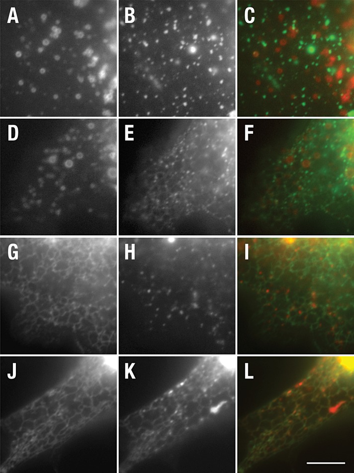
FIGURE 5:
CD8-NΔE LLAA accumulates in CD-MPR–positive tubular endosomes. PEN1/2 −/− MEFs were cotransfected with plasmid encoding SARA-mChr (A and D) and CD8-NΔE-GFP (B) or CD8-NΔE LLAA-GFP (E). Alternatively, cells were cotransfected with CD-MPR-GFP (G and J) and CD8-NΔE-mChr (H) or CD8-NΔE LLAA-mChr (K). Live cells were imaged after a 10-h incubation to limit recombinant protein expression levels. Merged images are shown in (C), (F), (I), and (L). Scale bar: 10 μm.

