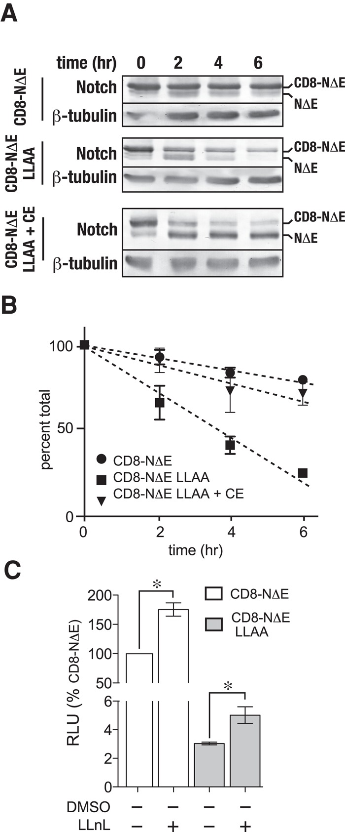
FIGURE 7:

γ-secretase cleaves activated Notch forms to generate proteosome-sensitive cleavage products within the endosome. (A) tTA HeLa cells were infected with adenovirus encoding either pCMV-CD8-NΔE or pCMV-CD8-NΔE LLAA in the presence or absence of 1 μM CE. Following a 16-h incubation, protein synthesis was inhibited by treating cells with cycloheximide, and Notch chimera stability was evaluated by immunoblotting at the indicated times; a representative blot is shown. (B) Densitometric analysis of Notch chimera stability from three independent experiments. In the plot, total Notch was determined by combining densitometric values of the upper and lower Notch bands. (C) CD8-NΔE– or CD8-NΔE LLAA–expressing tTA HeLa cells were treated with either dimethyl sulfoxide (DMSO) or LLnL for 6 h to disrupt the proteosome. Notch signaling was then evaluated using the luciferase assay. Error bars indicate ± SEM of three independent experiments; p values by t test: *, p < 0.05.
