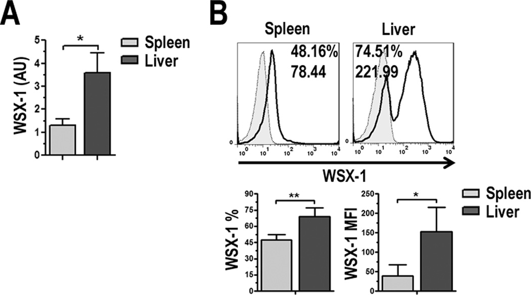
FIGURE 3.
pDC express the IL-27Rα/WSX-1. A, WSX-1 mRNA was determined by semi-quantitative RT-PCR for liver and spleen pDC and WSX-1 gene expression relative to β-actin was calculated. Data are expressed as AU; B, Liver and spleen pDC were stained for expression of IL-27Rα/WSX-1 and analyzed by flow cytometry. Values indicate percent positive cells and relative MFI compared to isotype controls (gray-filled histograms). Percentage of WSX-1 positive cells and MFI were averaged from 3 independent experiments, * p < 0.05, ** p < 0.01.

