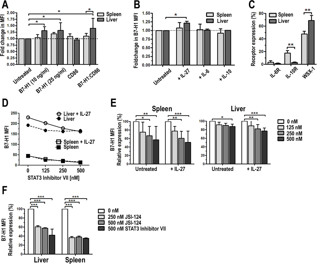
FIGURE 4.
IL-27 augments B7-H1 expression in liver pDC. PDCA-1-purified liver or spleen pDC were cultured for 18 h in the presence of 10 or 25 ng/ml IL-27. Cells were harvested, stained, and the expression of B7-H1 and CD86 examined by flow cytometry. Results are presented as fold change in B7-H1 MFI on IL-27-conditioned cells compared with untreated cells (set to 1.0); B, Liver pDC were cultured with 25 ng/ml IL-27, IL-6, or IL-10 for 18 h and B7-H1 expression was analyzed. Results represent fold change in B7-H1 MFI; C, Surface expression of the IL-6Rα, IL-10Rα and WSX-1/IL-27Rα were analyzed on liver and spleen pDC. Data represent percent positive cells, D, Experiments in A were repeated in the absence or presence of increasing concentrations of a STAT3 inhibitor (STAT3 Inhibitor VII). Data represent 4 independent experiments; E, Percent change in B7-H1 expression in the presence of the STAT3 inhibitor compared to untreated cells was calculated and averaged from 4 independent experiments; F, Experiments in D and E were repeated with a second STAT3 inhibitor JSI-124/Cucurbitacin at the indicated concentrations and compared to STAT3 Inhibitor VII. Results represent percent change in B7-H1 MFI in the presence of the STAT3 inhibitors and are an average of two independent experiments, * p < 0.05, ** p < 0.01, *** p < 0.001.

