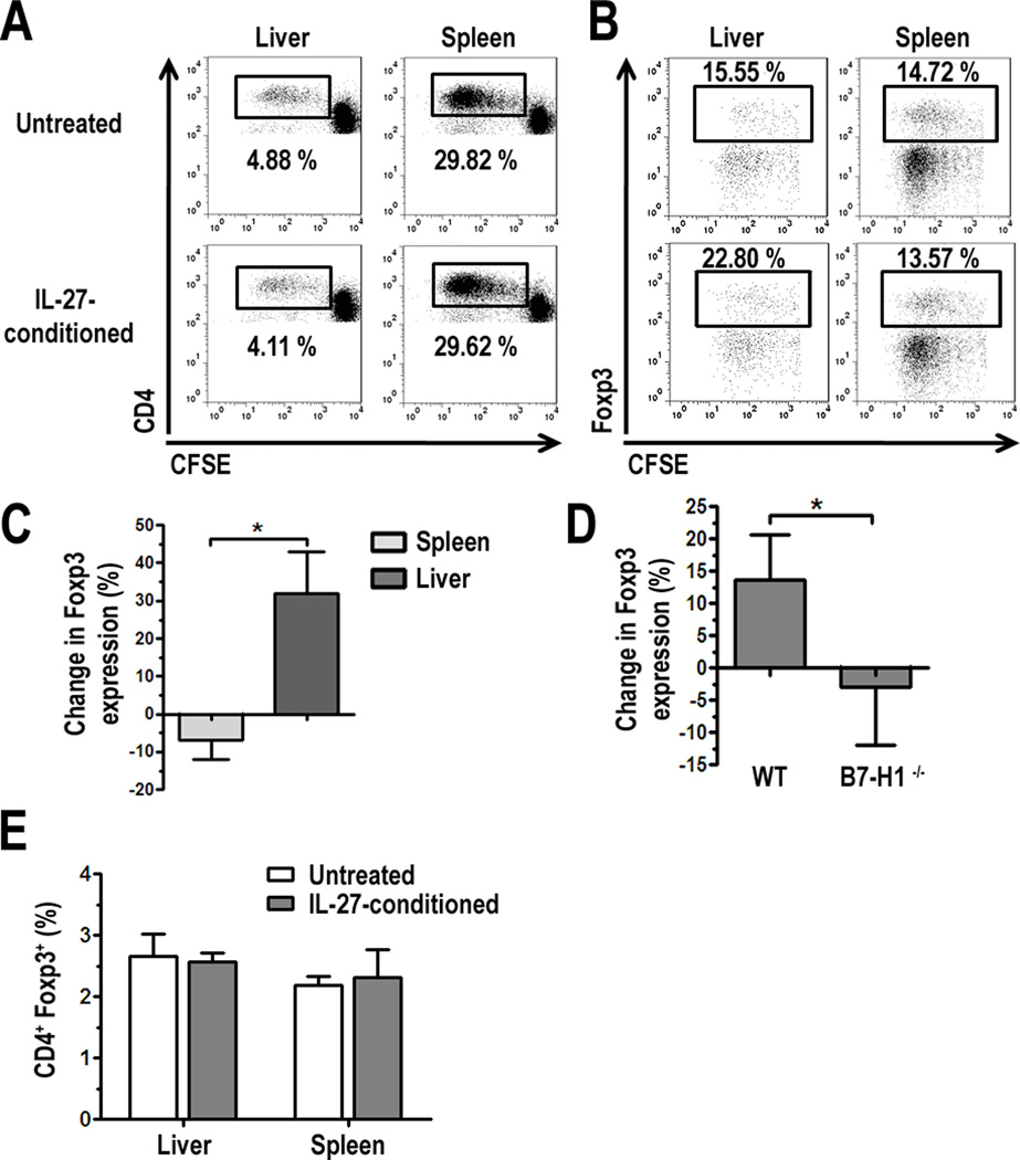
FIGURE 5.
IL-27-conditioned liver pDC increase the percentage of CD4+Foxp3+ T cells in MLR. A, Freshly-isolated liver or spleen PDCA-1+ pDC were cultured for 18 h with 25 ng/ml IL-27, washed, counted, and cultured in MLR with CFSE-labeled BALB/c splenic T cells. After 3 d, cells were harvested and stained for analysis of CD4+ T cell proliferation (A) and expression of Foxp3 (B) by flow cytometry. Data represents 3 independent experiments for both A and B; C, The percent change in Foxp3 expression in proliferating CD4+ T cells (cells gated in A) in cultures with IL-27-conditioned liver or spleen pDC compared to untreated pDC was calculated and average across 3 independent experiments; D, WT or B7-H1−/− liver pDC were cultured with IL-27 for 18 h and subsequently cultured with BALB/c T cells. Intracellular expression of Foxp3 was analyzed by flow cytometry after 3 d MLR and results were averaged across 3 independent experiments; E, Foxp3 expression was quantified in non-dividing CD4+ cells (CD4+ CFSEhi cells) from experiments shown in A, and averaged across 3 independent experiments, * p < 0.05.

