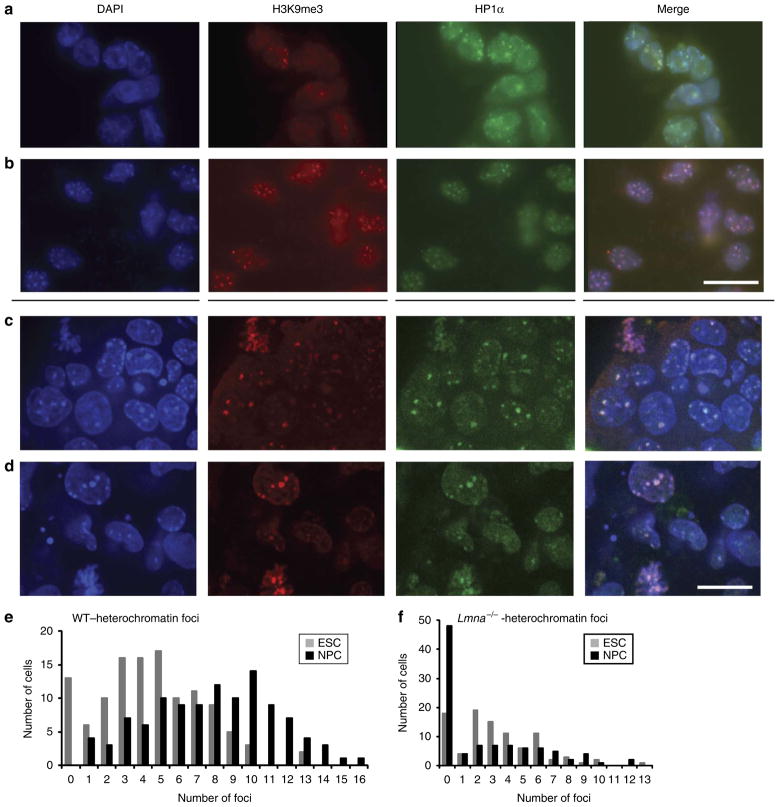
Figure 6. Lmna −/− NPCs exhibit abnormal heterochromatin structure.
(a,b) DAPI (blue) and immunostaining for heterochromatin markers H3K9me3 (red) and HP1α (green) in WT mouse ESCs (a) and NPCs (b). Scale bar, 20μm. (c,d) DAPI (blue) and immunostaining for heterochromatin markers H3K9me3 (red) and HP1α (green) in Lmna−/− ESCs (c) and in Lmna−/− NPCs (d). Scale bar, 20μm. (e) Distribution of heterochromatin foci number per nucleus in WT ESCs (grey) and NPCs (black). The average number of foci per nucleus increased from 4.5 ± 5.9 in ESCs to 7.9 ± 4.1 in NPCs. (f) Distribution of heterochromatin foci number per nucleus in Lmna−/− ESCs (grey) and NPCs (black). The average number of foci per nucleus decreased from 3.3 ± 6.8 in ESCs to 2.6 ± 12.1 in NPCs. At least 100 cells were counted in 2 independent experiments.

