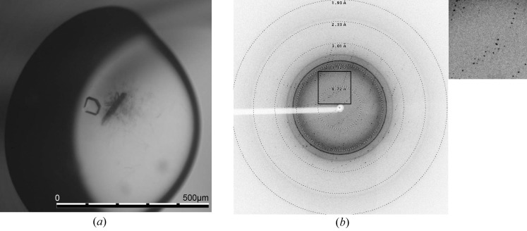
Figure 6.
TraMΔ crystallization and data collection. (a) A representative TraMΔ crystal, with compact growth with a size of less than 100 µm. The crystal was grown using the microbatch method at 293 K and with paraffin oil for sealing the plate. The protein drop ratio was 35% with a protein stock concentration of 3.0 mg ml−1. The drop size was 2 µl with the following final conditions derived from Index condition No. 44: 16.5% PEG 3350, 0.1 M HEPES, pH 7.33. (b) Diffraction pattern of a TraMΔ selenomethionine crystal obtained using synchrotron radiation on beamline X06DA, SLS, Villigen, Switzerland; resolution rings have been added. The picture was generated using ADXV (A. Arvail). Inset, detail of the diffraction shown in (b).

