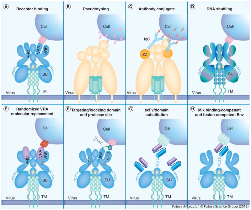
Figure 1. Approaches to alter retroviral receptor usage.
(A) Entry of a gammaretrovirus. The envelope (Env) protein consists of a trimer of the SU and TM proteins. The VRA and VRB function in receptor binding. The host cell receptor is depicted as a multiple transmembrane protein (Table 1). (B) Viral pseudotyping. The Env protein from an alternative virus (schematically depicted as sindbis virus) can associate with retroviral particles. The pseudotyped Env will bind to its cognate receptor, which is not limited to multiple transmembrane proteins [36–40]. (C) Antibody conjugation systems. Antibodies can be nonspecifically bound to modified Env proteins to direct viral entry. For sindbis virus, the mutation of the receptor-binding domains and insertion of the protein A ZZ domain allows for association with IgG molecules. Antibody binding to antigen delivers viral particles to cells [59–61]. (D) DNA shuffling. Combinations of related viral species (schematically shown using a six-color gradient) are mixed during PCR, allowing for the generation of complex chimeras derived from mixed portions of all of the parental sequences. These chimeras are then selected for properties including altered receptor recognition or protein stability [93]. (E) Randomized VRA or molecular replacements. For feline leukemia virus, substitutions within the VRA region are known to alter receptor usage. By randomizing 11 amino acids within the VRA, libraries of random Env proteins are generated and have been screened for functional entry into cells. The cognate receptors need to be identified. This method has identified novel Env/receptor pairs [14,26,27,98–100,103]. Alternatively, through the use of molecular modeling, specific substrates have been engineered into the VRA region of murine leukemia virus, allowing entry through the somatostatin receptor [84]. (F) Insertion of additional domains to either target or block the wild-type Env receptor-binding domain. For blocking domains, the cleavage by a host cell protease results in the release of the virus in the vicinity of the targeted cell [85–89]. Additional binding domains function to bind alternative receptors, but can allow the wild-type Env to function as a trigger for membrane fusion. (G) Domain substitutions. Binding domains can be used to substitute for a large section of the surface subunit protein. Examples of these types of substitutions include single-chain antibodies [32,51–55]. (H) Complementation studies. Viral entry involves more than receptor binding and requires a complex series of conformational changes to allow for membrane fusion. Viruses capable of binding, but not fusion, can be complemented with alternative fusogenic Env proteins [57,81,82].
SU: Surface subunit; TM: Transmembrane subunit; VRA: Variable region A; VRB: Variable region B; VRC: Variable region C; ZZ: IgG binding domain.

