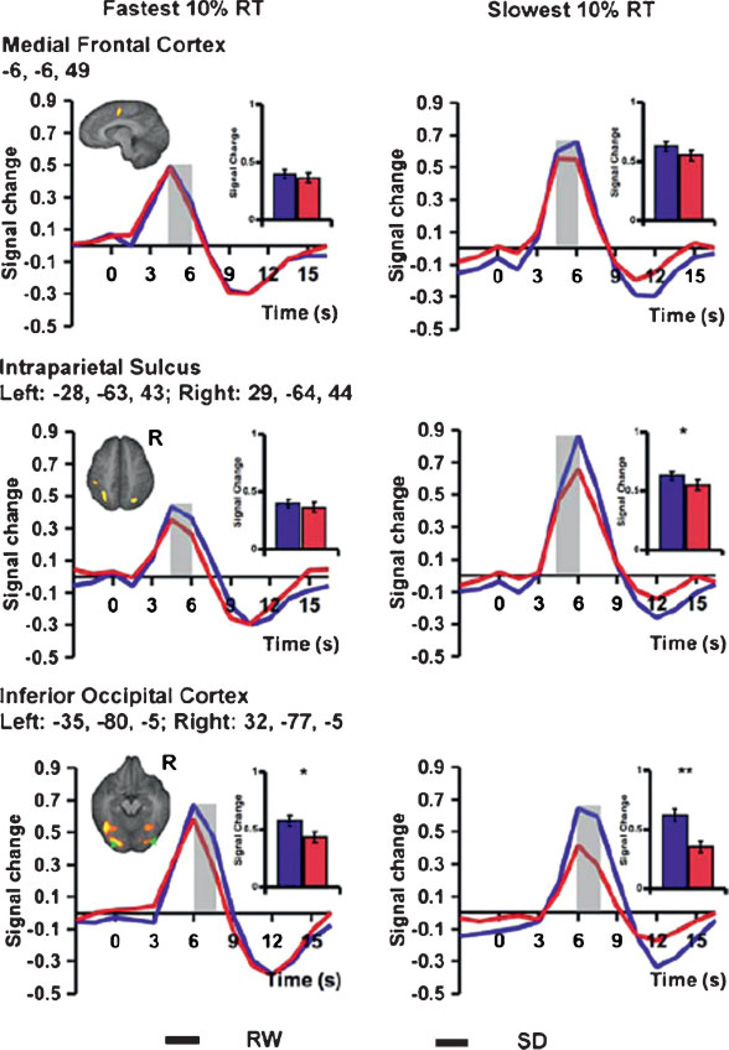
Figure 1.
Functional magnetic resonance imaging (fMRI) responses from three cortical areas during a visual, global/local selective attention task performed by N = 24 healthy young adults when not sleep deprived (RW, in blue) and when sleep deprived (SD, in red) for one night. The graphs display differential neural responses in the medial frontal cortex (top), bilateral intraparietal sulcus (middle), and bilateral inferior occipital cortices (bottom), in association with the fastest 10% reaction times (RT) (left column) and the slowest 10% RTs (right column). A threshold of p < 0.001 was used to detect task-related activation. For both RW and SD states, slower responses were associated with higher peak fMRI signals in the medial frontal cortex and bilateral intraparietal sulcus (all p < 0.005). When comparing SD with RW, peak signal for the slowest 10% of trials was significantly lower in the parietal and occipital regions (right middle and bottom), but not in the medial frontal cortex (right top). SD also attenuated task-related thalamic activation (not shown). Peak signal in the occipital region after SD was significantly lower than RW even for the fastest 10% of trials (left bottom). However, there was no difference between RW and SD states in the frontal or parietal peak fMRI signals for the fastest responses across states (left top and middle). The shaded time points indicate those contrasted to assess significant state effects. The inset shows the mean peak signal associated with the time points under consideration. Error bars represent SEM. Significant differences between peak signals associated with a lapse and the average response for each state are marked with an asterisk. *p < 0.01, **p < 0.001. (Reprinted from Chee et al,141 with permission from The Journal of Neuroscience.)

