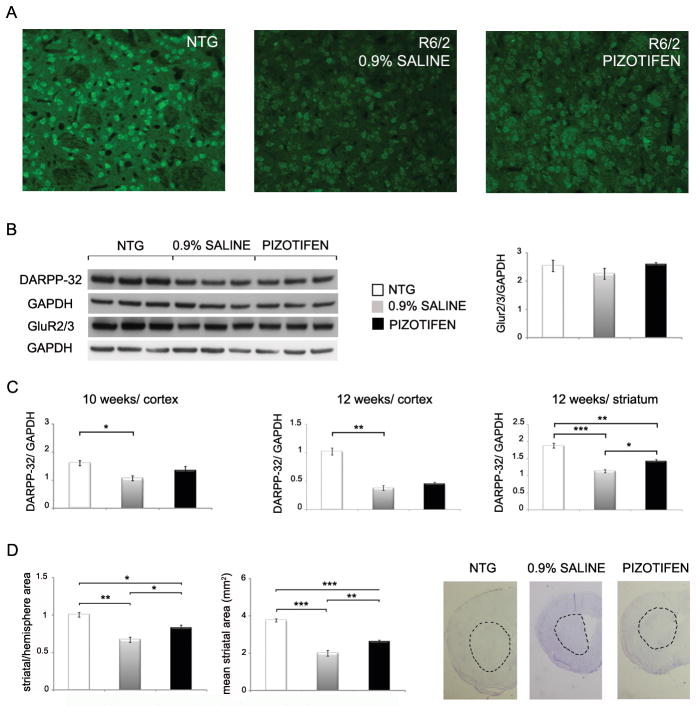
Figure 8. DARPP-32 expression and striatal area are improved in the pizotifen-treated HD transgenic R6/2 mouse model.
(A) Immunohistochemistry of DARPP-32 in the striatum of NTG, R6/2 (saline) and R6/2 (pizotifen) mice (n=3 mice, representative image shown). (B, left panel) Western blotting of DARPP-32 and mGlu2/3 AMPA receptors in the cortex in wild-type, R6/2 (saline) and R6/2 (pizotifen) mice (n=3 mice mean±SEM). (B, right panel) Densitometry analysis of GluR2/3 protein levels normalized to GAPDH in the cortex of NTG, R6/2 and pizotifen treated mice. (C) Side by side comparison of densitometry analysis of DARPP-32 protein levels in the striatum (12 weeks old) and cortex (10 and 12 weeks old) of age matched NTG, R6/2 and pizotifen treated mice. (D, left panel) Striatal area of the NTG, R6/2 (saline) and R6/2 (pizotifen) mice. (Right panel) Examples of sections analyzed. Demarcation shows striatal area as it was measured. * p<0.05, ** p<0.005, *** p<0.0005, 1-way ANOVA (n=3 mice mean±SEM).

