FIG. 1.
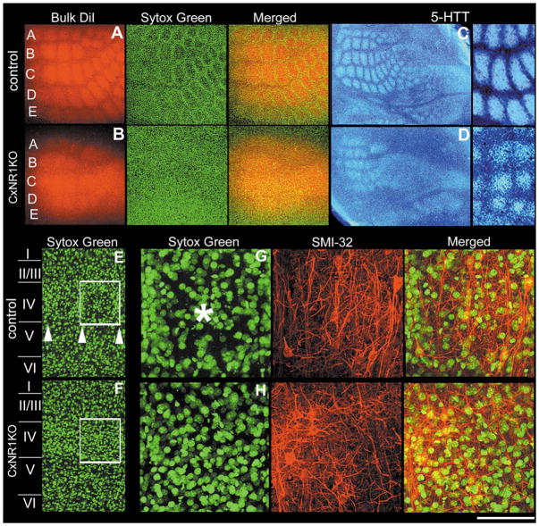
Layer IV cells in CxNR1KO S1 fail to organize into barrels while TCAs form rudimentary patterns. Series of micrographs of the same section and field in A illustrate the patterning of TCAs (red) and barrel cell nuclei (green) in the tangential plane in control mice. Similar series of photomicrographs in B illustrate the barrel cortex in the CxNR1KO mice in the same plane. C and D show inverted photomicrographs of TCA patterns visualized by anti-5-HTT immunohistochemistry in the tangential plane of the barrel cortex of control and CxNR1KO mice, respectively. Higher power images to the right show 5-HTT-immunopositive TCA regions versus the septal regions (quantified in Fig. 2). E and F show low-power views of layer IV and a single barrel in control (E) and uniform nucleoarchitecture in CxNR1KO (F) cortices. Series of micrographs in G and H are high-power views of a single barrel region depicted in the boxed regions in E and F, respectively. These series show the same region with different filters and merged images after staining with Sytox green for nuclei and SMI-32 for neuropil staining. Arrowheads in E mark the lower boundary of layer IV barrels and asterisk in G marks the center of a single barrel hollow which is absent in the CxNR1KO S1 cortex (H). Scale bar A, B = 400 μm; C, D = 750 μm; high power of C, D, and E, F = 150 μm; G, H = 50 μm.

