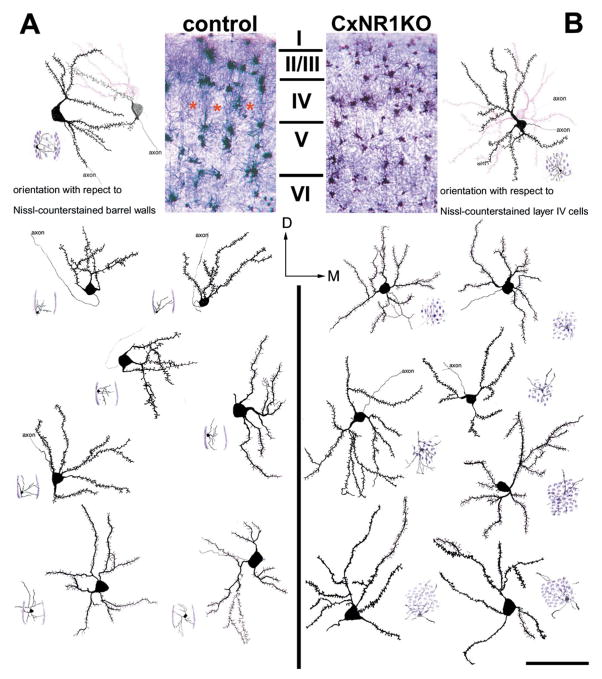
FIG. 5.
Layer IV spiny stellate cells in CxNR1KO fail to exhibit orientation bias. Low-power photomicrographs of Golgi and Nissi-stained coronal sections illustrate the cortical layers and the barrel field in control (A) and CxNR1KO (B) mice. In each panel multiple examples of camera lucida drawings of spiny stellate cells are shown. Small cartoon diagrams located in the bottom corner of each cell depict the location of the cell with respect to a single barrel in the control cases (A) and within a uniform distribution of Layer IV cells in the CxNR1KO cortex. Note that all of the cells located in the barrel walls of the control cases have biased dendritic tree orientations, while none of the cells charted in the CxNR1KO cortex show this bias. Scale bar A, B = 250 μm; inset = 80 μm.

