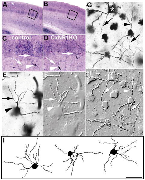
FIG. 7.
Aspiny layer IV cells in the CxNR1KO and control S1 cortex. Low-power photomicrographs of NADPH-stained coronal sections show distinct patterns in layer IV of the control barrel cortex (A) and less distinct patterns in the CxNR1KO cortex (B). Higher magnification of the boxed areas in micrographs A and B show a few aspiny stellate cells positive for NADPH (C, D). These cells (arrowheads) are often seen below or above layer IV with their axonal processes (arrows) extending into layer IV. (E, G) show two Golgi-stained aspiny stellate cells and photomontage-embossed images (F, H) from the layer IV of P33 control and CxNR1KO cortex, respectively. Arrowheads in E and F point to the cell body and the arrows to the same aspiny dendritic branch. In G and H, arrows point out to the same axonal process while arrowheads point to the aspiny dendritic branch. Note the spiny stellate cell (asterisk) just above and to the left of the aspiny cell. I illustrates camera lucida drawings of aspiny stellate cells from a P33 CxNR1KO barrel cortex. A, B = 600 μm; C, D = 150 μm; E, F = 30 μm; G, H = 40 μm; and I = 60 μm.

