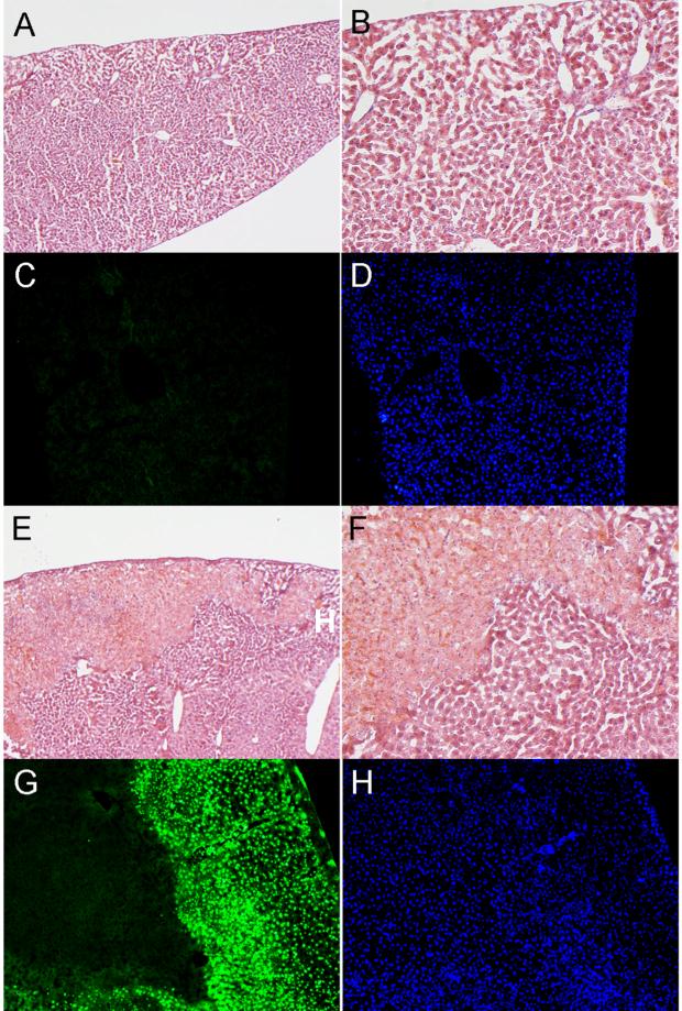
Figure 4.
In vivo biocompatibility of PAGS. Mice were either injected with 8 mg of PAGS or 0.2 mg of PEI intraperitoneally. Of all the organs isolated, liver was the only one that showed any significant histological change. A representative image of liver tissue in the PAGS group, H&E staining (A, 100x and B, 200x) revealed normal tissue architecture and TUNEL (C, green fluorescence) revealed no apoptosis. (D) DAPI staining revealed nuclei in the same tissue section as C. For PEI at 0.2 mg/animal, H&E staining (E, 100x and F, 200x) revealed that liver tissue suffered approximately 15% necrosis. (G) TUNEL staining showed extensive apoptosis in the livers of the PEI groups. (H) DAPI staining revealed nuclei in the same tissue section as G. All images were from samples isolated day 1 post injection. All image acquisition parameters are identical for PAGS and PEI.

