Figure 2.
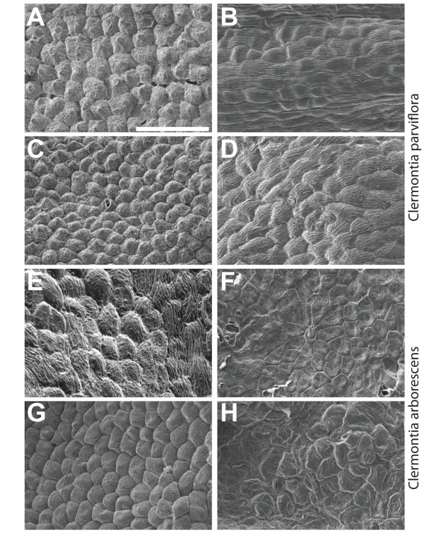
Epidermal micromorphology of perianth organs. Scanning electron microscope images from (A-D) C. parviflora and (E-H) C. arborescens on the adaxial (left panels) and abaxial (right panels) epidermal surfaces. Canonical Conical epidermal cell morphology, an indicator of petal identity, is discovered are apparent on the adaxial surface of the outer perianth organs of C. parviflora (A). Similar cone-shaped cells are observed on the adaxial surface of inner perianth organs in both C. parviflora (C) and C. arborescens (G). However, the outer perianth organs of C. arborescens display a flattened morphology more typical of sepal organs (E). Scale bar denotes 100 μm.

