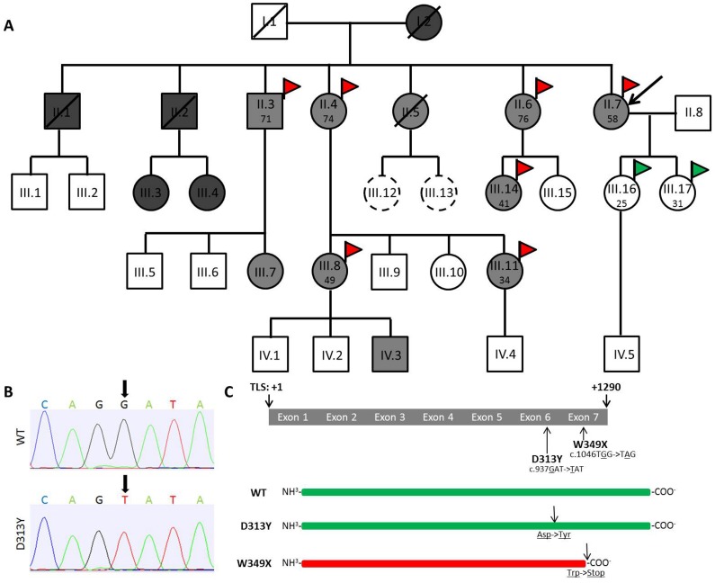
Figure 1. Pedigree and positions of D313Y and W349X in the GLA coding region.
(A) Pedigree. (B) Representative chromatograms showing nucleotide substitution at position +937 (G>T) in the GLA coding region. (C) Schematic overview of the GLA transcript including localizations of D313Y and W349X. The pedigree shows the complete family of index patient II.7. Black arrow in A labels index patient. Squares indicate males, circles indicate females. Diagonal lines indicate deceased family members. Dark grey, light grey, and white color in squares and circles indicates W349X, D313Y, and non-carriers, respectively. Scattered circles represent not sequenced patients. Red flags indicate patients with white matter lesions (WML) seen in magnetic resonance imaging (MRI), green flags indicate control patients without WML. Patient’s age at MRI is given in years. D313Y results in single amino acid substitution at position +313, leading to a conversion of aspartate (Asp) to tyrosine (Tyr). W349X results in a c-terminal truncated GLA protein, due to a conversion of tryptophan (Trp) to a stop-codon. A: Adenine; C: Cytosine; G: Guanine; T: Thymine; TLS: translational start side; WT: wild-type GLA without any coding mutations.

