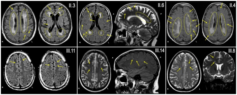
Figure 3. Punctuated (arrows) and confluent (yellow circles) WML were present on MR images (axial and sagittal FLAIR- and T2-sequences) of all six examined family members, all carrying D313Y.
Only patient II.3 had mild cardiovascular risk factors (treated arterial hypertension). Extent and lesion load were age-related, but WML were already present in young family members without any vascular risk factor (patient III.8, III.11 and III.14; 49, 34 and 41 years of age, respectively).

