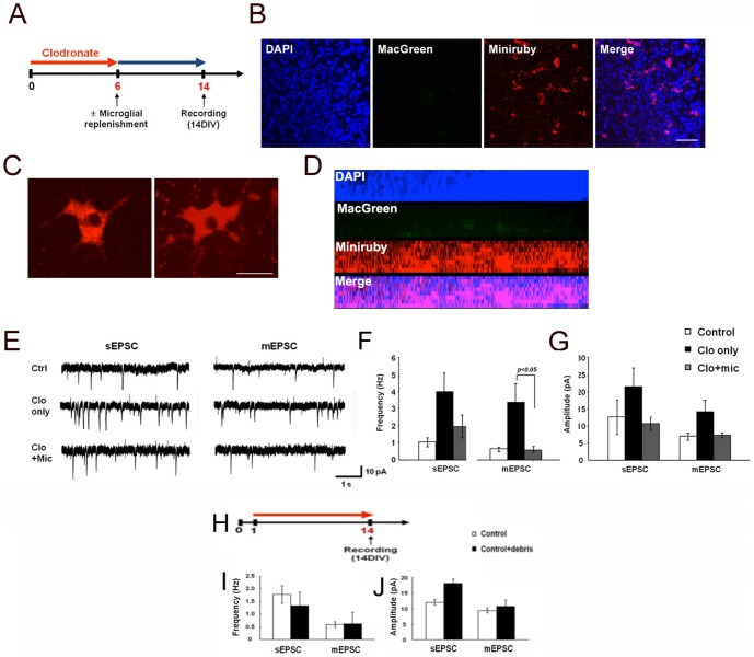
Figure 3. Replenishment of microglia reverses the effect of microglial depletion on organotypic brain slices.
A. Organotypic slices obtained from MacGreen mice were treated with clodronate for 6 DIV. Clodronate was washed away and then the slices were cultured for 8 DIV in the absence of clodronate with or without primary microglia labeled with miniruby (5 µg/ml, 8×10² cells in 3 µl). B–D. Images showing exogenously added microglia (Miniruby, red) to CA1 hippocampal slices treated with clodronate. MacGreen indicates endogenous microglia (green). Higher magnification of miniruby+ microglia in C. Side view of the z-stacking images for B. DAPI staining was used to visualize nuclei. Scale bars, 50 µm in B; 20 µm in C. E–G. Recording traces (E) and summaries of frequency (F) and amplitude (G) of sEPSCs and mEPSCs from CA1 neurons in control, clodronate-treated, and clodronate-treated with replenished microglia organotypic slices. Error bars represent mean ± SEM. H–J. Cellular debris does not affect sEPSC and mEPSC frequency or amplitude. H. Organotypic slices were incubated either without (control) or with (red line) added cellular debris (control+debris). I, J. Summary of sEPSC and mEPSC frequency (I) and amplitude (J) from control or cultures with added cellular debris at DIV14. Data are expressed as means ± SEM (n = 2 for each).

