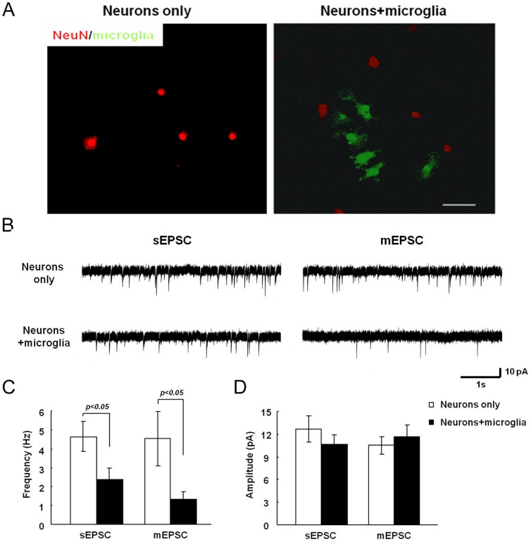
Figure 4. Addition of microglia to neurons reduces mEPSC frequency.
Primary hippocampal neurons (DIV21) were cultured for 2 days either alone or in the presence of microglia obtained from MacGreen mice. A. Neurons were stained with anti-NeuN antibody and visualized with Alexa Fluor555-conjugated secondary antibody. Scale bar, 50 µm. B. mEPSC recordings from neurons in the presence or absence of microglia. C, D. Summary of mean mEPSC frequency (C) and amplitude (D) from neurons in the presence or absence of microglia.

