Figure 2.

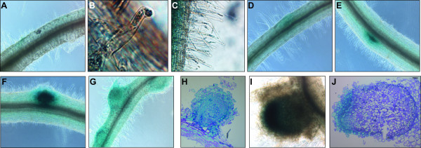
MtN5 promoter activity during rhizobial infection and nodule development. Representative expression patterns are shown. Localization of MtN5 expression in the root epidermis (A) and in the root hairs (B, C) 3 hours post-inoculation (hpi). (D) GUS staining detected in the root cortex at 24 hpi. MtN5 promoter activity in nodule primordia (E, F) and in young nodules (G, H). MtN5 promoter activity in fully developed root nodules (I, J).
