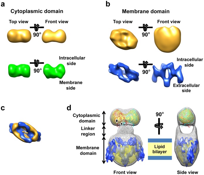
Figure 3. Fitting of the atomic structure of cytoplasmic domain.
[14] and the 7.5 Å resolution 2D crystal structure of membrane domain [23] into our 3D map of full length AE1 dimer. (a) Shaded surface views of the atomic structure of cytoplasmic domain (PDB ID: 1HYN) filtered to 2.4 nm resolution (green) compared to the corresponding views of cytoplasmic domain resolved in the EM single-particle reconstruction (gold) of full-length AE1 dimer. In the EM map, the membrane domain of AE1 dimer is removed for clarity. The two structures are similar in size and in having a double-humped shape on their cytoplasmic side. (b) Shaded surface views of AE1 membrane domain resolved from 2D crystals embedded in trehalose (EMDB ID: 1645) filtered to 2.4 nm resolution (blue), as compared to the corresponding views of membrane domain resolved in the EM single-particle reconstruction (gold). The extracellular and intracellular sides identified in the published 2D crystal structure were used to define the orientation for comparison. (c) Superposition of the two structures of membrane domains described in (b) viewed from the cytoplasmic side (top view). The EM single-particle reconstruction is rendered at higher density threshold to show the deep canyon, which is consistent with the membrane domain structure from 2D crystals. (d) Fitting the EM single-particle reconstruction of full-length AE1 dimer with the crystal structure of cytoplasmic domain (red and cyan) and 2D crystal structure of membrane domain (blue). The single-particle reconstruction is rendered in two density threshold values: at low threshold (gray mesh) and a high threshold (yellow). The approximate positions of N-terminus and C-terminus of the cytoplasmic domain are labeled with diamond and triangle, respectively.

