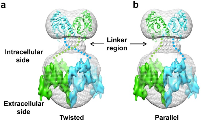
Figure 4. Two possible modes of monomer-monomer association in AE1 dimer.
(a) The twisted mode of dimerization. (b) The parallel mode of dimerization. The crystal structure of cytoplasmic domain (ribbon) and 2D crystal structure of membrane domain (surface) are fitted into the single-particle reconstruction of full-length AE1 dimer (gray mesh) and the two monomers (each consisting of a cytoplasmic domain and a membrane domain) are colored in green or cyan, respectively. The tentative linkers connecting cytoplasmic and membrane domains are depicted as broken lines.

