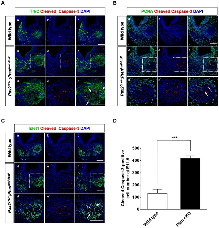
Figure 5. Neuronal apoptosis in the cochleovestibular ganglion (CVG) complex of Pten-deficient mice.
(A–D) Apoptotic cells in the CVG were stained with anti-cleaved caspase-3 antibody at E11.5. Cleaved caspase-3 immunoreactivity (red) increased substantially in Pten-deficient mice compared to that in wild-type mice. (A) Apoptotic neurons were occasionally co-localized with TrkC-positive (green) (arrows in f) or negative cells. Higher magnification images of d, é, and f are shown in the insets in d, e, and f, respectively. Scale bars: 100 µm. (B) Proliferating PCNA-positive cells (green) were not seen in apoptotic neurons (arrows in f ´). Higher magnification images of d, e, and f are shown in the insets in d, e, and f, respectively. Scale bars: 100 µm. (C) Cleaved caspase-3-positive cells (red) were also Islet1-negative cells (arrows in f ´). Apoptotic neurons (red) were distributed in the core of the non-proliferative area that expressed Islet1 (green). Higher magnification images of d, e, and f are shown in the insets in d, e, and f, respectively. Scale bars: 100 µm. (D) Numbers of cleaved caspase-3-positive apoptotic cells in the CVG were significantly increased in Pten-deficient mice at E11.5 (3 cochleae, P<0.001).

