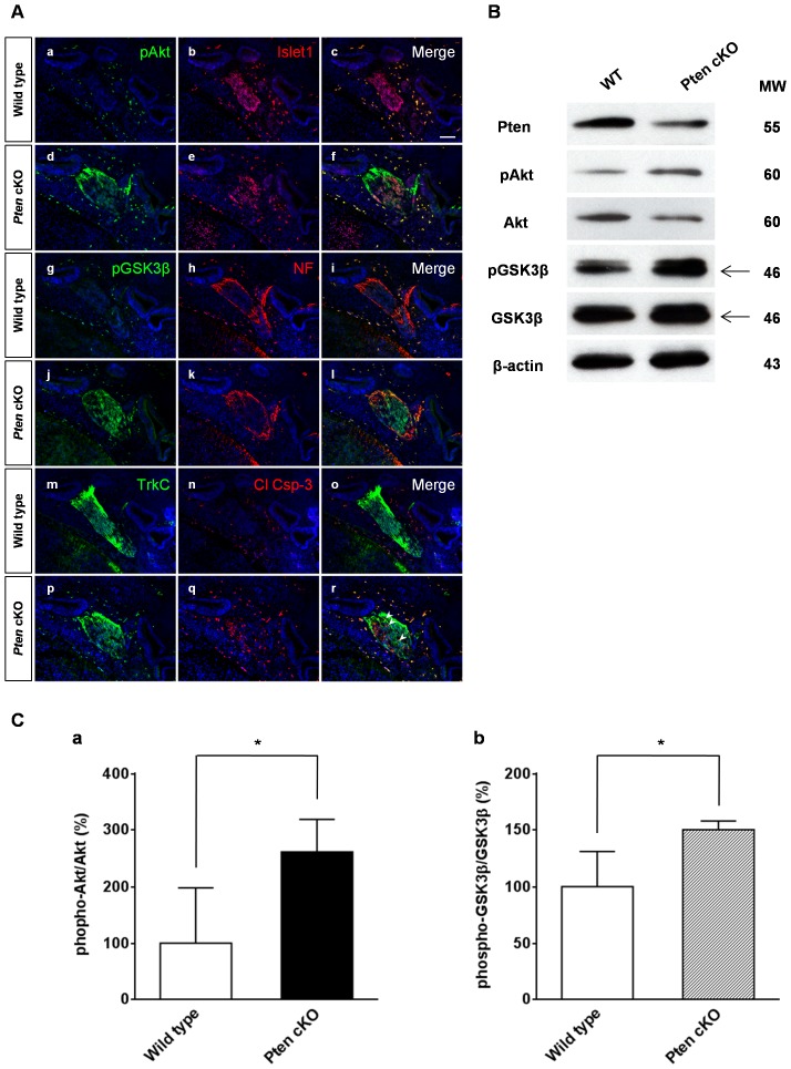
Figure 6. Akt and GSK3β phosphorylation in Pten-deficient mice.
(A) Phosphorylation of Akt and GSK3β was measured by immunofluorescence staining using anti-phospho-Akt (Ser473) (pAkt) and anti-phospho-GSK3β (Ser9) (pGSK3β) antibodies at E13.5. Compared to wild-type spiral ganglia, Akt phosphorylation (green) increased substantially in Pten-deficient mice (a–f). GSK3β phosphorylation (green) was also highly expressed in the spiral ganglia of Pten-deficient mice compared to that in wild-type mice (g–l). In the absence of Pten, all pAkt and pGSK3β cells were maintained in Islet-1 positive cells (red) (a–l). Wild-type mice showed well-organized patterns of Islet-1-positive cells (a–c), whereas Pten-deficient mice showed a slightly scattered form of spiral ganglia (d–f). Cleaved caspase-3-positive apoptotic neurons (red) were sometimes co-localized with TrkC-positive cells (green) (arrowheads in r). DAPI-stained nuclei (blue) are seen in all images. Scale bar: 100 µm. (B) Western blotting analysis of Pten, pAkt, Akt, pGSK3β, GSK3β, and β-actin expression in the inner ear at E14.5. Proteins were extracted from four pooled inner ears at E14.5. (C) The relative intensity of each phospho-protein was normalized to the total level of the same protein. Levels of both pAkt (a) and pGSK3β (b) were significantly increased in Pten cKO mice (4 cochleae, P<0.05).

