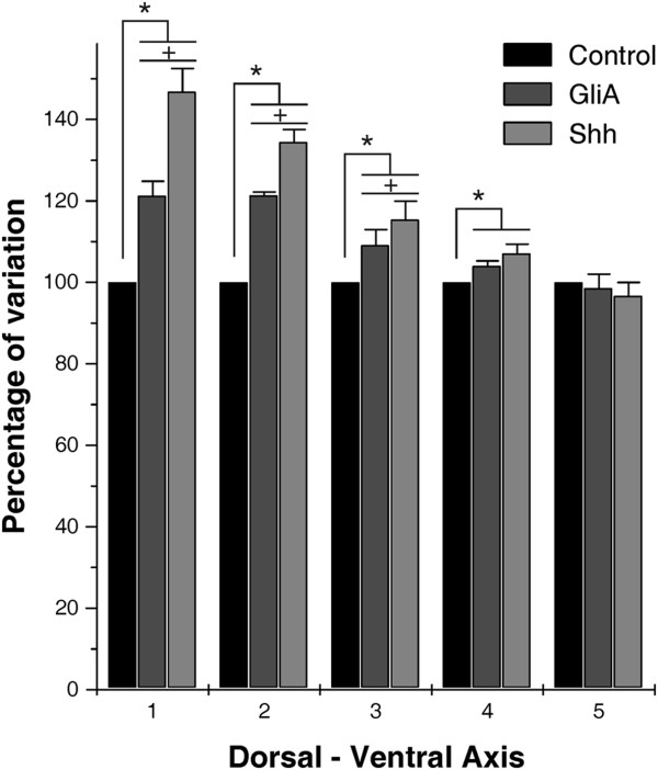
Figure 8.
Space-dependent differences of Shh and GliA effects along the D-V axis. Each bar represents the mean ± standard deviation of the mNEc density measured in 500 μm length spatial windows located at defined positions along the D-V axis. The means mNEc density measured in controls OT were taken as reference (100%). A decreasing responsiveness to Shh and GliA can be observed from the dorsal region (1) to the ventral one (5). 1: dorsal region, 3: halfway between the OT dorsal and ventral zone; 5: OT-tegmental boundary. 2 and 4 represents intermediate positions between 1 and 3 and between 3 and 5 respectively. *: indicate statistically significant differences (p<0.001) measured by the Z test. +: indicate statistically significant differences (p<0.01) between GliA and Shh.

