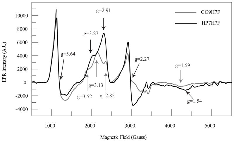
Figure 5.
X-band EPR comparison of ferric HP7-H7F and CC9-H7F. As expected based on the relatively small KA,his, both spectra contain a mixture of high- and low-spin Fe(III) complexes. The signal near g= 6 is similar to that of myoglobin and other 5 ferric heme examples containing an axially coordinated histidyl imidazole ligand. The rhombic signals with features in the range from g = 3.52 to 1.54 are typical of bis-histidine coordinated low-spin Fe(III) complexes.

