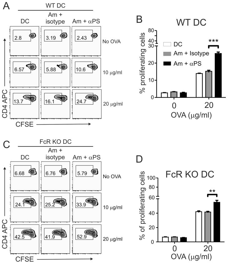
Figure 4. Blocking PS recognition enhances overall antigen presentation capacity of infected DCs.
(A, B) C57BL/6 WT and (C, D) FcR KO BMDCs were infected with lesion-derived amastigotes that were pre-treated with 9 μg/ml of mAb mch1N11 (PS-targeting) or isotype antibody in the presence or absence of different concentrations of OVA protein. After 24 h of infection, DCs were harvested and co-cultured with CFSE-labeled OTII CD4+ T cells for 3 days. The percentage of proliferating T cells was determined by CFSE dilution. (A, C) Graphs are representative of 3 independent experiments. (B, D) Graphs represent data from 3 pooled experiments. ** p <0.01, *** p <0.001.

