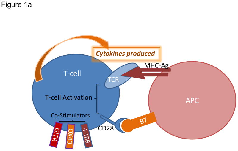
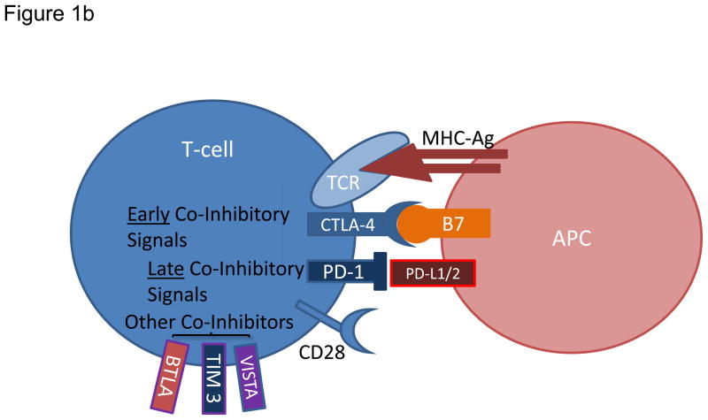
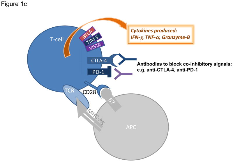
Figure 1. Modulation of T cell activation and current strategies promoting effector T cell functions.
a) Augmenting T cell activation by positive co-stimulation. Antigenic presentation triggers T cell activation and occurs when a peptide bound to major histocompatibility complex (MHC) molecule on an antigen presenting cell (APC) interacts with T cell receptor (TCR) on the surface of a T cell. In order to achieve optimal activation, additional co-stimulatory signals are required and primarily involve interaction between CD28 on T cells and B7 on APCs. Other T cell positive co-stimulators include 4-1BB, OX40, and GITR. b) Limiting T cell activation by negative co-stimulation. After T cell activation, cytotoxic T lymphocyte-associated protein 4 (CTLA-4) is mobilized to the cell surface and binds to B7 with greater affinity than CD28 therefore preventing signaling through CD28. Later inhibitory signals can be provided by co-inhibitors such as programmed cell death 1 (PD-1), which binds to PD-1 ligand 1 (PDL1). Other co-inhibitors of T cell activation include VISTA, TIM3, and BTLA. c) Sustaining T cell activation through blockade of negative co-stimulatory molecules. Blocking antibodies against CTLA-4 or PD-1 are currently employed to neutralize co-inhibitory receptors and prevent dampening of the T cell response. Blockade of these inhibitory immune checkpoints results in enhanced and sustained activation of tumor-specific T cells that produce cytokines including tumor necrosis factor-α (TNF-α), interferon-γ (IFN-γ) and granzyme B.


