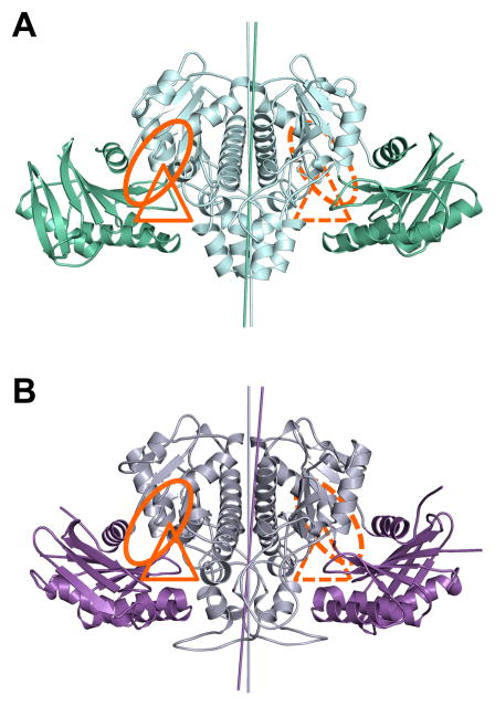
Figure 2. Structures of the eukaryotic ACKs.
A) E. histolytica ACK with the N-terminal wing domain colored green and the C-terminal body domain colored cyan. B) C. neoformans ACK with the wing domain colored purple and the body domain colored grey. The putative acetate and nucleotide binding sites are highlighted with a triangle and a circle, respectively. The rotation axes relating each domain of the dimer are highlighted with a line colored similarly to the corresponding domain.

