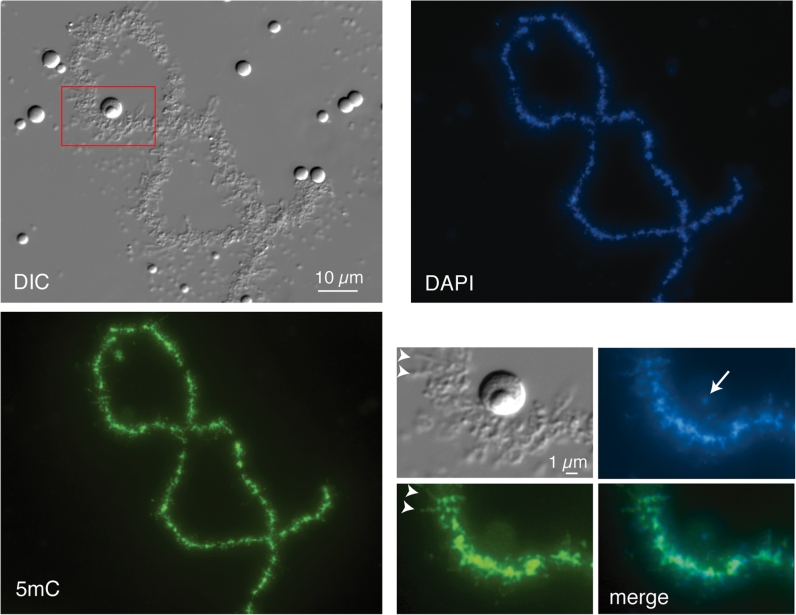
Fig. 5.
Immunostaining of Xenopus laevis LBC with 5-methylcytosine mAb. At lower magnifications a general chromomeric staining for 5mC is evident that is proportional to the DNA concentration indicated by DAPI staining. The region shown at higher magnification in the insets is indicated by the red box in the DIC image. Arrowheads in the insets indicate two of the 5mC-stained fibrils that project laterally from the chromomeric axis and that presumably correspond to the bases of some lateral loops. Note that the amplified rDNA that can be specifically detected in the fibrillar centres of extrachromosomal nucleoli by DAPI staining (arrow) appears unstained for 5mC, consistent with the lack of methylation in amplified rDNA determined by biochemical analyses (Dawid et al. 1970)

