
Figure 3.
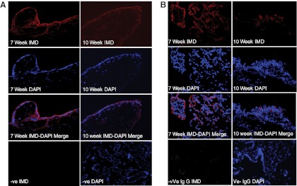
Expression of IMD immunoreactivity in first-trimester placental villi and decidua. A, IMD immunoreactivity in electively aborted villous tissues collected from 7 and 10 weeks of gestation (n = 3). B, IMD immunolocalization in decidua collected from 7 and 10 weeks of gestation (n = 3). IgG was used as negative control and DAPI was used for blue nuclear staining (×400 magnification). DAPI, 4′,6′-diamino-2-phenylindole.