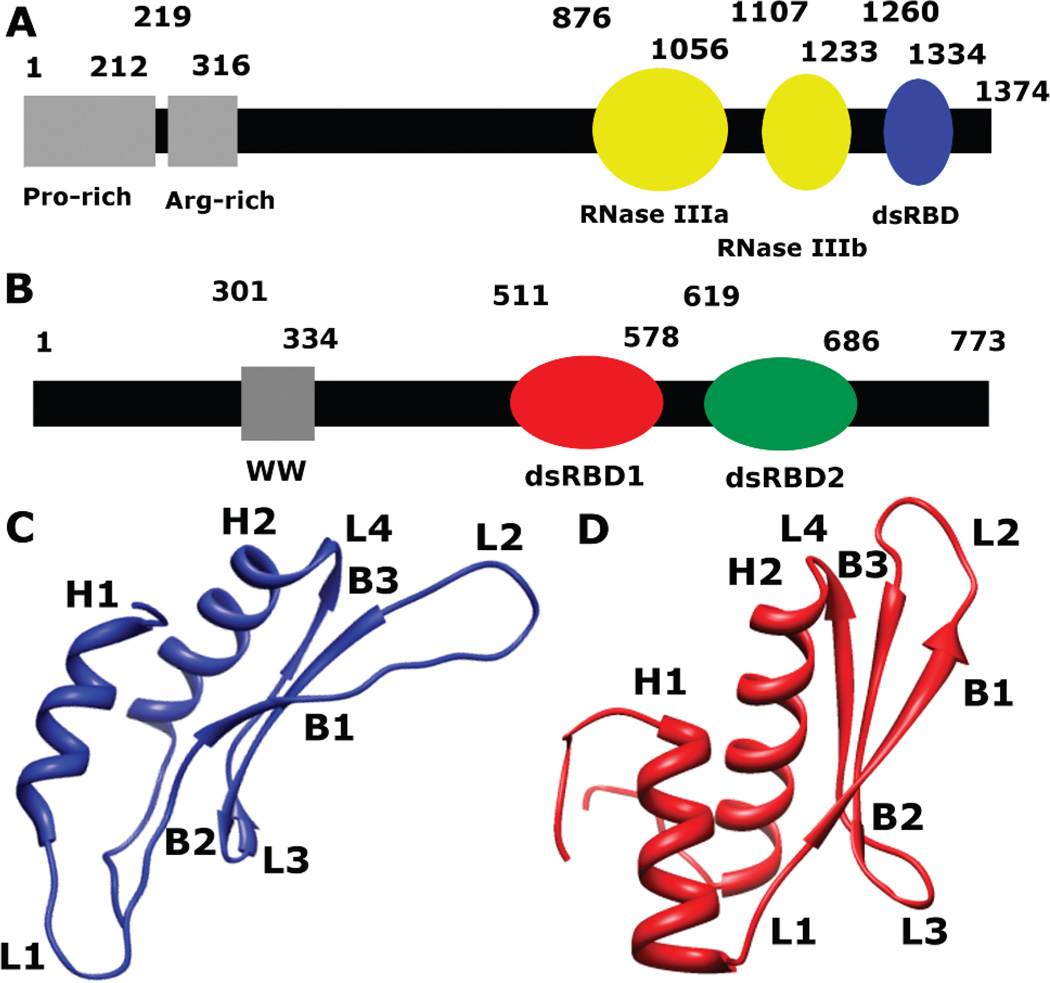
Figure 2.
Schematic representation of the primary sequence of (A) Drosha and (B) DGCR8. (C) A ribbon diagram representing the solution structure of Drosha-dsRBD (PDB 2KHX, residues 1259–1337) shows the extended loops 1 (L1) and 2 (L2). (D) A ribbon diagram representing the crystal structure of DGCR8-dsRBD1 (PDB 2YT4, residues 505–583) shows a less elongated fold of the dsRBD than Drosha-dsRBD.

