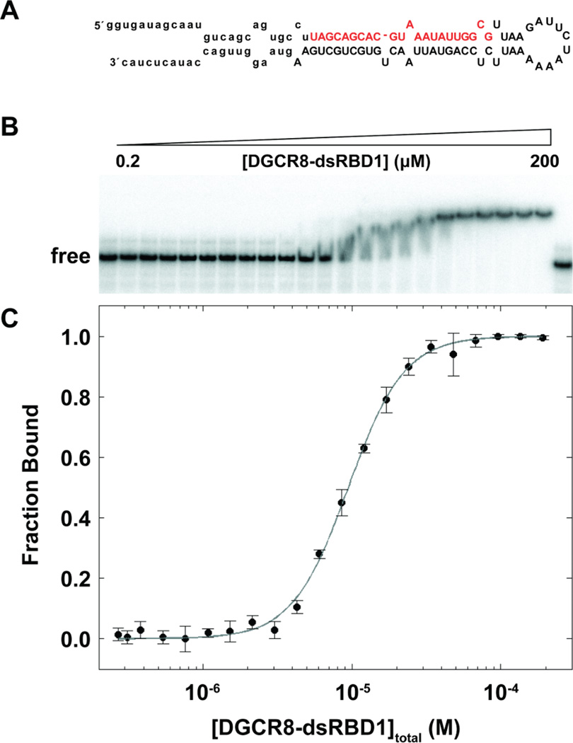
Figure 3.
EMSA of pri-miR-16-1 binding to DGCR8-dsRBD1. (A) Predicted secondary structure of pri-miR-16-1 with the sequence of the mature miRNA shown in red and the region removed by Drosha cleavage indicated through lower case letters. (B) Representative gel showing addition of DGCR8-dsRBD1 (2–200 µM) to 0.25 nM pri-miR-16-1. (C) Fitted EMSA fraction bound as a function of DGCR8-dsRBD1 concentration with data points and uncertainties represented by filled circles and the best fit to the data (see text) represented as a gray line.

