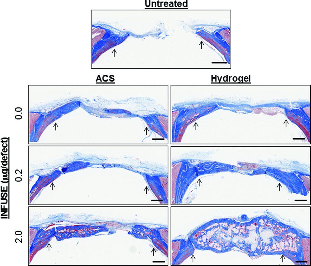
Figure 4.
Histological evaluation of bone regeneration. Coronal tissue sections through bone defect sites were prepared from de-calcified skulls taken upon completion of the study. Representative sections stained with Masson’s Trichrome are shown with arrows pointing to original defect edge. Additional histology images are presented in Supplemental Figure 3. Scale bars represent 1mm.

