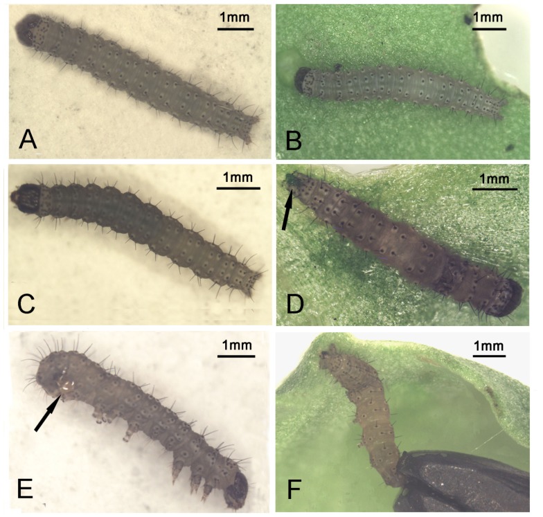
Figure 2.
(A, B) Healthy larvae with light and uniform body color; (C) A darker patch appeared anteriorly on a dying larva poisoned by cantharidin; (D) Darker patches spread all over the body of a dead larva poisoned by cantharidin with wet, green frass stuck to its anal area, as shown at the arrow; (E) Mucus was kept between the fourth pair of prolegs and caudal prolegs as shown at the arrow; (F) A larva that died from cantharidin was glued posteriorly and ventrally to a leaf by mucus.

