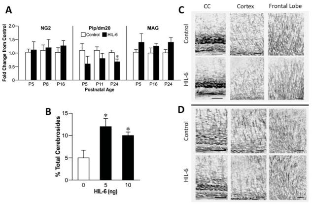
Figure 5.
(A) qRT-PCR for myelin specific transcripts in the cortex at various postnatal days (P) relevant to the developmental profile of each: NG2 (P5, 8, 16), PLP/DM20 (P5, 11, 24), and MAG (P5, 16, 24) of mice injected at P4 with vehicle (white bars) or 5ng HIL-6 (dark bars). ANOVA indicated a main effect of HIL-6 for PLP/DM20 and MAG. *significant post-hoc analysis with Bonferroni correction for multiple comparisons (p=0.014) as compared to vehicle. (B) NFA cerebrosides as a percent of total cerebrosides (NFA-cerebroside fraction) in adult mice injected at PND4 with either vehicle or HIL-6 (5 ng). Data represents the mean % value ± SEM. *significantly different from controls (p<0.05).
(C–D) Representative photomicrographs of immunostaining for myelin basic protein (MBP, 1:500, 20hr, 4°C) as detected by NBT/BCIP. Staining intensity and complexity of MBP myelin tracts increased with age (C: PND16 [Suppl. Fig. 5]; D: PND24). Branch points observed in the HIL-6 mice suggested less complexity of myelinated fibers.

