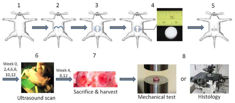
Fig. 1.

Study design. 30 female 16-wk old Lewis rats under anesthesia were implanted with the 60 scaffolds (20 scaffolds per type with 2 matched type scaffolds per animal). Scaffolds were implanted in the rat’s abdomen from the midline incision, with each sample sutured into a full thickness circular abdominal wall defect surgically created in the left or right side. The week 0 scan was performed 3 days after surgery and the scans afterwards were performed bi-weekly from the first scan day until sacrifice at week 4 (n=3, one on each group), week 8 (n=3, one on each group) and week 12 (n=4, one on each group). Tissue constructs were harvested for compression testing or histological staining.
