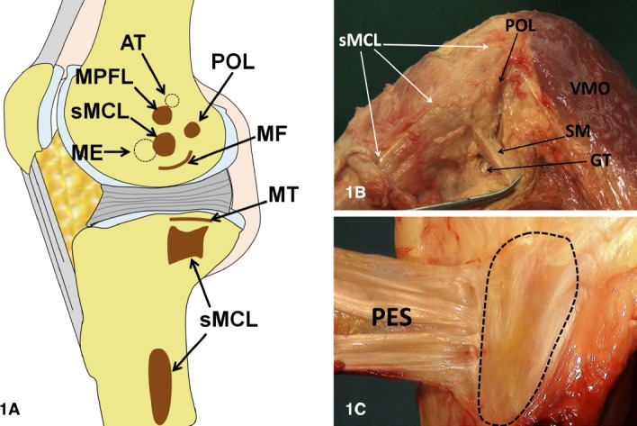
Figure 1. A) Medial view of the knee: attachment sites (Redrawn from: laprade Rf et al. The anatomy of the medial part of the knee. J Bone Joint Surg Am. 2007;89(9):2000–10). B) Medial aspect of the knee. C) sMCl distal tibial insertion. AT= adductor tubercle, ME= medial epicondyle, sMCl= superficial medial collateral ligament, Mpfl= medial patellofemoral ligament, and pOl= posterior oblique ligament. Mf= Meniscofemoral, MT= Meniscotibial, SM= Semimembranosus tendon, vMO= vastus medialis obliquus, GT= Gastrocnemius tendon, pES= pes Anserinus.

