
Figure 3. 75%/25% Histological Analysis.
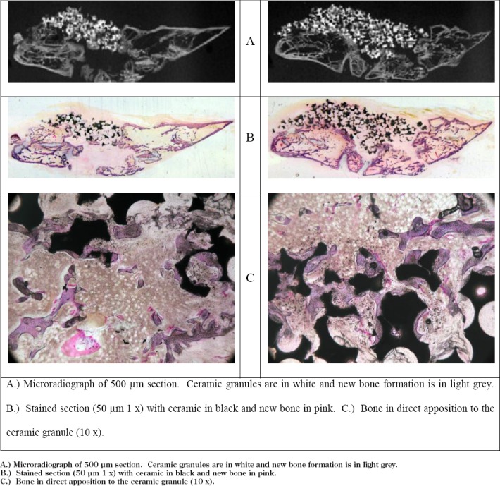
A.) Microradiograph of 500 μm section. Ceramic granules are in white and new bone formation is in light grey.
B.) Stained section (50 μm 1 x) with ceramic in black and new bone in pink.
C.) Bone in direct apposition to the ceramic granule (10 x).