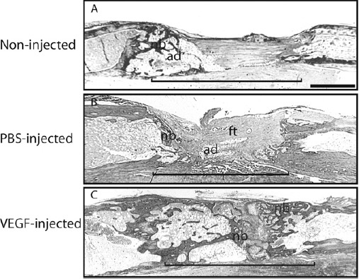
Figure 2. Histological analysis at 27 days after completing rapid distraction. Tissue is stained by trichrome staining. (A) A non-injected distraction gap exhibits a small amount of new bone (nb) at the osteotomy ends and adipose (ad) and fibrous tissues (ft) in the distraction gap (bracket). (B) Similar histological findings are observed in a saline-injected distraction gap. (C) A VEGF-treated distraction gap is bridged by new bone (nb). Recanalization of the regenerated bone is also seen in VEGF injected animals. ad = adipose tissue, ft = fibrous tissue, nb = new bone, bracket = distraction gap. scale bar = 1mm.