Figure 6.
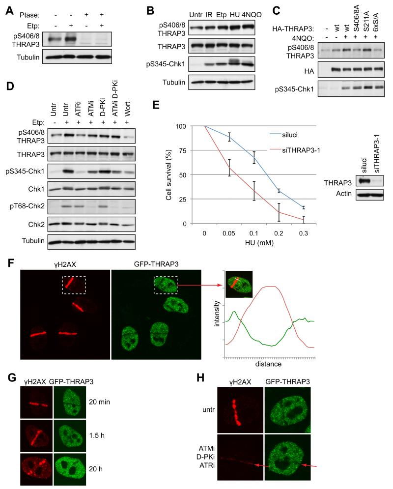
THRAP3 is phosphorylated in response to DNA damage and is excluded from DNA damage sites. (A) and (B) Phosphorylation of THRAP3 in cell extracts untreated or treated with etoposide (Etp; 3 μM, 2h), HU or 4NQO, which were treated with λ phosphatase (Ptase) where indicated. (C) Phosphorylation of wild-type HA-THRAP3 or indicated THRAP3 phospho-mutants in U2OS cells treated with 4NQO where indicated. (Note that antibody detection was not reduced further by mutating all 6 phosphorylation sites to Ala; 6×S/A). (D) Phosphorylation of THRAP3 in cells treated with inhibitors of ATM (ATMi), DNA-PK (D-PKi), ATR (ATRi) or wortmannin (Wort) for 1 hour, then treated with Etp (3 μM, 2h). (E) Clonogenic survival of cells transfected with siluci or siTHRAP3-1 and treated with HU at the indicated doses. Results are presented as an average from 3 experiments −/+ SEM. Depletion of THRAP3 is shown by Western blotting. (F) Exclusion of THRAP3 from sites of laser-induced DNA damage in HeLa-GFP-THRAP3 cells. Quantification of green (GFP-THRAP3) and red (γH2AX) was performed by Volocity software. The graph shows the intensity of fluorescence (y-axis) along the white line (x-axis) indicated by arrow. (G) Exclusion of THRAP3 from laser-damage in cells fixed at indicated times. (H) Exclusion of THRAP3 from DNA damage sites is inhibited in cells treated with inhibitors of ATM (ATMi), DNA-PK (D-PKi) and ATR (ATRi) for 1 hour prior to micro-irradiation. (untr = untreated; p = phospho). (See also Figure S6)

