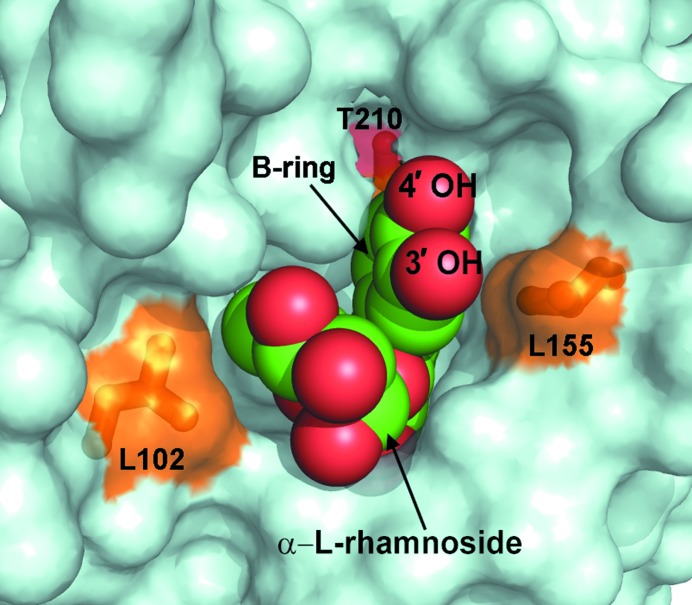
Figure 5.
The quercitrin molecule nested in the binding pocket between the N- and C-lobes of mRSK2NTKD. Quercitrin is shown using van der Waals spheres (oxygen in red and carbon in green); the protein is depicted as a surface representation. The three key residues making close contacts with quercitrin (and also depicted in Fig. 6 ▶) are shown as sticks and are colored and labeled.

