
Figure 6.
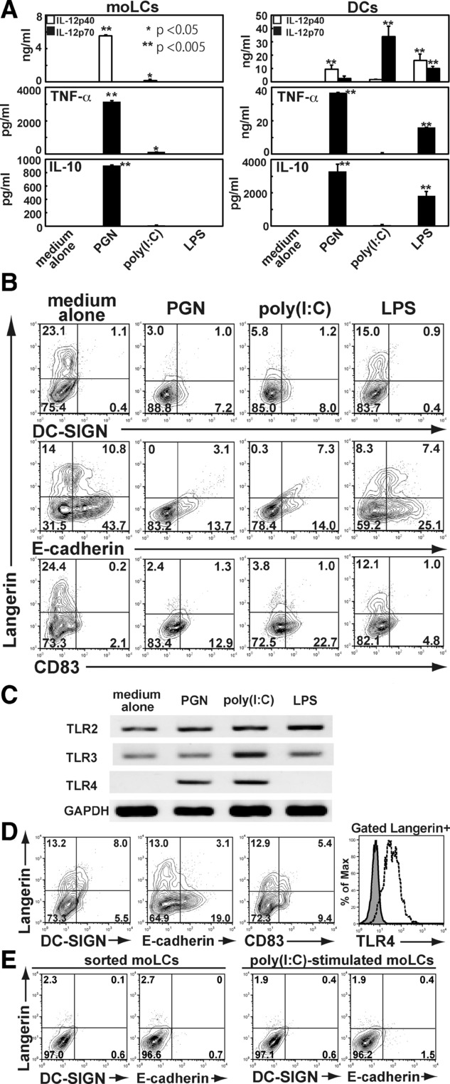
PBMo-derived, moLCs irreversibly became activated DCs via TLR–ligand stimuli. (A) PBMo-derived moLCs (left) or DCs (right) was stimulated with known TLR agonists; 20 μg/mL PGN for TLR2, 50 μg/mL poly(I:C) for TLR3, or 100 ng/mL LPS for TLR4, and cytokine production was measured by ELISA. Data are shown as the mean + SEM (n = 4) of results pooled from four independent experiments. *p < 0.05, **p < 0.005, Student's t-test . (B) PBMo-derived moLCs stimulated by those TLR-ligans for 24 h were analyzed their expression of Langerin, DC-SIGN, E-cadherin, and CD83. (C) Ligand-stimulated moLCs were examined for the expression of TLRs. (D) Langerin+ moLCs separated by sorting with magnetic cell separator (MACS) were analyzed their expression of DC-SIGN, E-cadherin, CD83, and TLR4. (E) Sorted moLCs (left) or poly(I:C)-stimulated moLCs (right) were stimulated on a human E-cadherin-coated plate and examined their expression of Langerin and E-cadherin. Results shown are representative of four independent experiments.