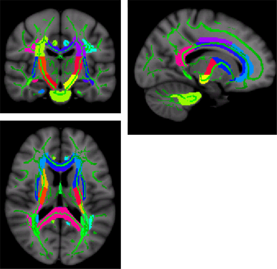
Figure 2.
“Skeletonized” DTI map indicating FA ROIs (colors) used to derive the summary FA score superimposed onto the mean FA skeleton (green) of the DTI subjects. The summary score comprised the following tracts: middle cerebellar peduncles, pontine crossing tracts part of the middle cerebellar peduncles, genu of the corpus callosum, body of the corpus callosum, splenium of the corpus callosum, fornix column and fornix body, corticospinal tract, medial lemniscus, inferior cerebellar peduncles, superior cerebellar peduncles, cerebral peduncles, anterior limb of the internal capsule, posterior limb of the internal capsule, retrolenticular part of the internal capsule, anterior corona radiata, superior corona radiata, posterior corona radiata, posterior thalamic radiation including optic radiation, sagittal striatum including the inferior longitudinal fasciculus, external capsule, cingulum-cingulate gyrus, cingulum hippocampus, fornix cres stria terminalis, superior longitudinal fasciculus, uncinate fasciculus, and the tapetum.

