Figure 1.

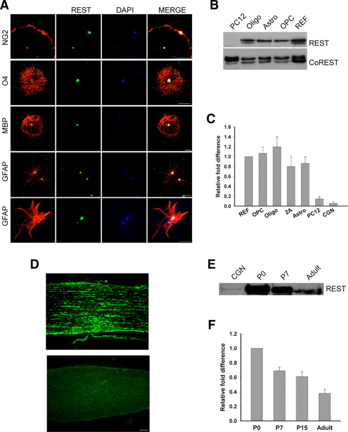
REST is expressed in the nuclear compartment of primary rat glia and in rat optic nerve. Purified OPCs, astrocytes, and oligodendrocytes were grown as described in Material and Methods for 5–6 d, and REST protein and transcript levels were measured. A, Immunofluorescence staining detects REST in the nuclei of cultured glial cells. Scale bar, 50 μm. B, Immunoblot showing the detection of REST and CoREST protein in nuclear extracts prepared from oligodendrocytes (Oligo), astrocytes (Astro), OPCs, and REFs. REST was not present in extracts of PC12 cells. C, Quantitative real-time PCR analysis showing the fold changes in REST mRNA in rat glia relative to REFs after normalization to GAPDH. REST expression is lower in PC12 cells and CGNs. Error bars represent the SD; n = 3. D, Immunofluorescence detection of REST in postnatal day 12 rat optic nerve (top). The bottom is control staining with secondary antibody alone. Scale bar, 50 μm. E, Immunoblot analysis of REST protein in P0, P7, and adult rat optic nerve. CGNs serve as a negative control. F, Real-time PCR analysis showing the fold change in REST expression in P7, P15, and adult rat optic nerve relative to P0 after normalization to GAPDH. Error bars represent the SD from six PCR runs; three runs each from two separate experiments.
