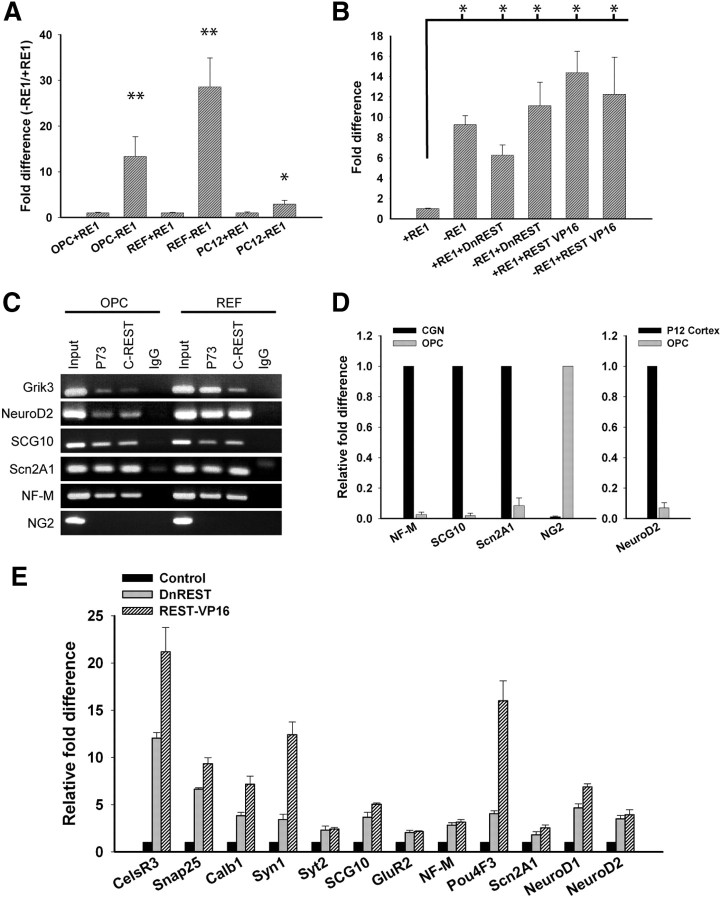
Figure 2.
REST is a functional repressor protein in OPCs. A, Luciferase reporter assay showing that RE1-containing reporters are repressed in OPCs but not PC12 cells. Error bars represent the SD from three separate experiments. *p < 0.03, **p < 0.01. B, Luciferase reporter assay showing that DnREST and REST–VP16 derepressed luciferase gene expression in the presence of an RE1. Error bars represent the SD; n = 3. *p < 0.01. C, ChiP assays showing that REST binds to an RE1 element in Grik3, NF-M, SCG10, Scn2a1, and NeuroD2 but not to a randomly chosen site upstream of the NG2 (CSPG4) gene in both OPCs and REFs. D, Quantitative real-time PCR showing the fold difference in NF-M, SCG10, and Scn2a1 relative to CGNs, NG2 relative to OPCs, and NeuroD2 relative to P12 rat cortex after normalization to GAPDH. Error bars represent the SD; n = 3. E, DnREST and REST–VP16 induced changes in the expression of RE1-containing genes. Purified OPCs were infected with DnREST, REST–VP16, or GFP control adenoviruses and grown in proliferation media for 72 h. Transcript levels of REST-regulated neuronal genes were analyzed by real-time PCR. The relative fold change in gene expression is shown relative to control-infected cells after normalization to GAPDH. Error bars represent the SD from six PCR runs; all gene expression changes had p < 0.01.

