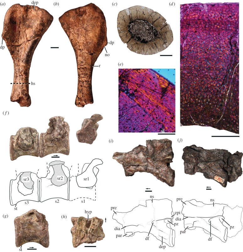
Figure 1.
Holotype (a–h; NHMUK R6856) and referred (i–j; SAM-PK-K10654) specimens of Nyasasaurus parringtoni gen. et sp. nov. Right humerus in (a) anterior and (b) posterior views. Histological section of humerus in (c) complete cross-section in transmitted light, (d) cross-section through the entire cortex in crossed Nicols, and (e) cross-section through the outer portion of cortex in crossed Nicols. (f) Rearticulated sacrum in right lateral view with interpretive drawing (below). (g) Posterior presacral vertebra in right lateral view. (h) Partial posterior presacral vertebra in dorsal view. (i) Anterior cervical vertebra in left lateral view with interpretive drawing (below). (j) Anterior cervical vertebra in left lateral view with interpretive drawing (below). Arrows point anteriorly. Scale bars, (a,b,f–j) 1 cm, (c) 4 mm, (d) 1 mm, (e) 500 nm. Dep, depression; df, deep fossa; dia, diapophysis; dp, deltopectoral crest; epi, epipophysis; hs, histology section; hyp, hypantrum; no, notch; ns, neural spine; par, parapophysis; pre, prezygapophysis; pz, postzygapophysis; r, ridge; s1–3, sacral vertebra number; sr1–3, sacral rib number; st, striations.

