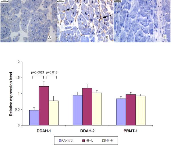
Figure 3. Increased Myocardial DDAH-1 Protein Levels in Human Failing Myocardium Caption: Upper Panel.
Upper Panel. Immunohistochemistry staining of DDAH-1 in donor hearts (A) and failing human hearts (B), as well as IgG control staining of failing human hearts (C). Arrows showing increased staining in the interstitial and perivascular areas. Lower Panel. Myocardial levels of DDAH-1/2 and PRMT-1 proteins among “Controls” (n=10), “HF-L” (“low” sPAP <50 mmHg, n=10) and “HF-H” groups (“high” sPAP ≥50 mmHg, n=10). Band densities were normalized to those of GAPDH.

