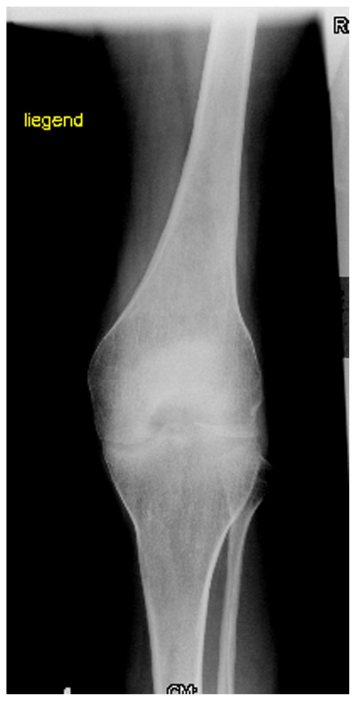
Figure 8.

AP radiograph of the knee showed severe osteoarthritis matching the grade of II–III of Kellgren-Lawrence grading scale. Note the flattened and dysplastic epiphyses associated with a marked narrowing of the joint spaces, sclerosis and subsequent deformity of the bony contour, the patient started to walk with crutches. The knee was unstable medial and lateral +++, patella was sub-luxated to the lateral and medial side, atrophy of the quadriceps muscle was evident, the MRI indicated an infarction-area in the distal femur.
